Development of Simulation and Prediction Models and Integration with the TCIA Database of Medical Images

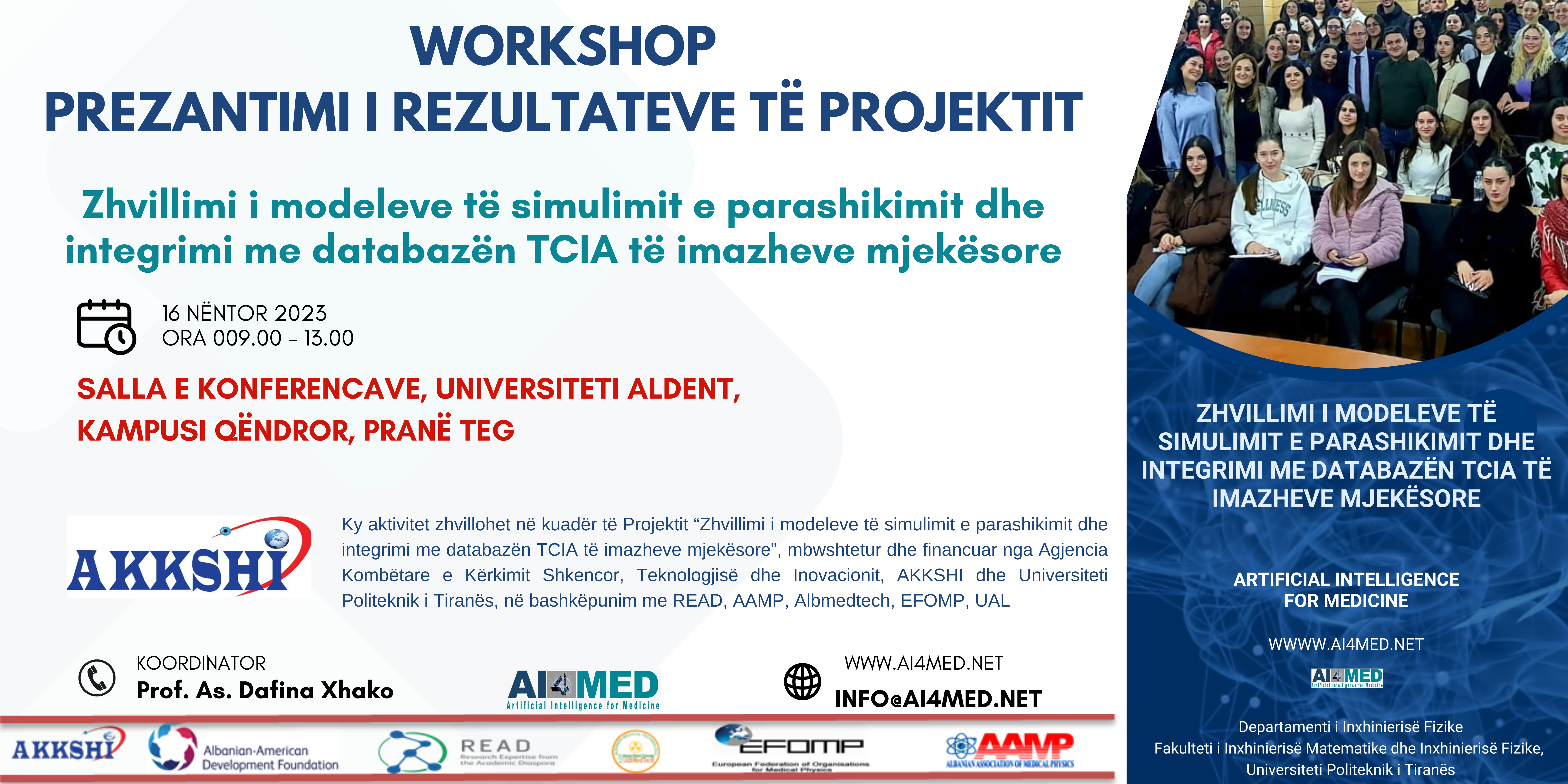
We are pleased to invite you to a special workshop dedicated to presenting the results of the project “Development of Simulation and Prediction Models and Integration with the TCIA Database of Medical Images.” This event will take place on:

📅 Date: November 16, 2023
🕘 Time: 09:00 – 13:00
📍 Location: Conference Hall, Aldent University, Central Campus (near TEG)

This workshop is organized within the framework of a project funded by the National Agency for Scientific Research and Innovation, and coordinated by Prof. As. Dafina Xhako. It brings together a network of collaborators including:

  • AKKSHI (Albanian American Development Foundation)
  • READ
  • AAMP (Albanian Association for Medical Physics)
  • EFOMP (European Federation of Organizations for Medical Physics)
  • UAL (University Aldent)

🔍 About the Workshop

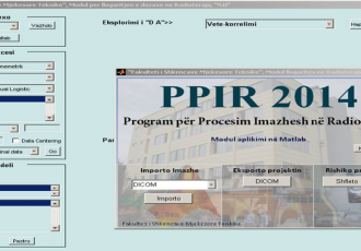
The event will showcase the development and integration of advanced simulation and prediction models with the TCIA (The Cancer Imaging Archive) database. These innovations aim to enhance the capabilities of medical imaging technologies, contributing to more accurate diagnostics and improved patient outcomes.

Participants will have the opportunity to:

  • Explore the technical and scientific achievements of the project
  • Engage with experts in medical physics, AI, and biomedical research
  • Network with professionals and institutions involved in cutting-edge medical imaging research

📩 Registration & Contact

To register or learn more, please visit: www.AI4MED.NET
For inquiries, contact us at: This email address is being protected from spambots. You need JavaScript enabled to view it.

Evaluation of quality radiologic devices using RTI evaluation method and comparing the results with international standards

Abstract:The diagnostic and radiological service, which is rated as a primary service, for the examination of internal diseases and the consequences of Covid-19, played a crucial role in examinations of patients with corona-virus. In this study, we aim to evaluate the important role that diagnostic imaging has played in dealing with the pandemic situation where, by means of X-ray imaging technologies, it has been possible to identify the severity of the disease in patients affected by Covid 19. With support of NASRI, under project "Development of simulation and forecasting models and integration with the TCIA database of medical images", we analysed the diagnostic system in several diagnostics centres in Albania. We used Piranha Multi (RTI Group) to verify the accuracy of the voltage (kV), the stability of the repetition of the values of the voltage dependence of the power voltage change, the overall filtering and the exposure time. We have presented a general picture of the situation of diagnostic equipment in Albania compared to OECD and COCIR indicators.  The study shows that diagnostic imaging in Albania uses a large variety of equipment, but compared to the OECD standard, the ratio of units per 1 million inhabitants in Albania is below the average number. We found a low level of compliance with the COCIR standards ("Golden Rules") where more than 65% of the basic equipment installed is between six and ten years old, 20% is less than five years old and 15% of the installed equipment is more than ten years old. 

Participation at the International Conference on Artificial Intelligence in Medical Application 8-9 June 2023, Hamburg, Germany

After several months of tiresome work, we are nearing the finalization of the project for the application of Artificial Intelligence in medical imaging. It is a privilege for our working group to present today some of the results of our work at the international conference: "International Conference on Artificial Intelligence in Medical Application 8-9 June 2023, Hamburg, Germany".
Thanks to the support of the National Agency for Scientific Research and Innovation #AKKSHI and Research Expertise from the Academic Diaspora #READ, we managed to get to know each other and become part of the network of professionals working in this direction! Our works: " Using Deep Convolutional Neural Network to create a DCNN model for brain tumor detection" and   "MRI image segmentation with HD Brain Extraction extension in 3D Slicer", were part of discussions with other collegaues.  New collaborations are comming: #artificialintelligence
#UPT, #FIMIF, #AKKSHI, #READ, #EFOMP

New method to calculate the tumor control probability for PPIR

Abstract: The methodology of using numerical methods in medical imaging and radiotherapy for calculations and simulations is very important. We are working for more than 10 years for implementing and developing several numerical methods in MATLAB. Using MATLAB, we can analyze data, develop algorithms, and create models and applications. We finalized the first Albanian module for training and simulation in radiotherapy called PPIR. One of the most important applications of this module is medical images processing, visualization and calculation in radiotherapy. The dose calculation on the PPIR is based on Eudmodel for dose volume histograms (DVH), which in radiotherapy treatment plan evaluation relies on an implicit estimation of the tumor normal tissue complication probability (NTCP) and control probability (TCP). In this work we present another model to determinate the tumor control probability (TCP) by analyzing the interactions of radiation with each tumor cell through virtual simulations. For a homogeneously irradiated tumor with a dose, our simulator determines the TCP using acceptable real-simulations of a fractioned treatment, based on the tumor cell sensitivities of radiation, tumor volume, cell density and number of fractions. We calculated the TCP from virtual simulations of a fractioned treatment as ratio of simulations with 100% of killed cells and total of simulations. The function is compared with Eudmodel and is incorporated with PPIR in a new version of PPIR2019

https://doi.org/10.1063/1.5135450

Seeing inside the human body through diagnostic imaging

Seeing inside the human body through diagnostic imaging is extraordinary. But even more impressive is the numerous possibilities of using artificial intelligence in determining the preliminary diagnosis for patients.
Every day we are taking small steps to understand as much as possible the capacities that artificial intelligence offers in diagnostics medicine. With the valuable contribution of Prof. Kita Sallabanda, we are working in the framework of the project project “Development of simulation and forecasting models and integration with the #cancerimagingarchive database of #medical images” 🩻. The main goal of the project is the use of artificial intelligence models in diagnostic imaging. With Prof.Kita and my colleague Niko Hyka, are currently determining the initial images as imputs and then we will continue with the configuration of the model, training of ANN and simulation process.
We are very pleased with this opportunity and support given by #AADF, Albanian - American Development Foundation (AADF),
#READ Research Expertise from the Academic Diaspora
Let's move forward
#opportunity #construction #project #artificialintelligence#diaspora#READ#albania#research#university #development #development #medicine #database#training

logo

Development of simulation and forecasting models and integration with the TCIA database of medical images